How to recognise and treat eczema in skin of colour

Delayed diagnosis and undertreatment of eczema in patients with skin of colour remain a persistent challenge in primary care. Here, Dr Abi Pararajasingam explains how to recognise the signs and deliver more equitable care.
Atopic eczema (atopic dermatitis) is one of the most common inflammatory skin conditions encountered in primary care and community pharmacy. Yet its recognition and management in patients with skin of colour remains an area where disparities persist. Differences in clinical presentation, gaps in training curriculums, and limitations in traditional severity scoring tools can all contribute to delayed diagnosis and undertreatment1,2.
For frontline clinicians, improving confidence in recognising eczema across diverse skin tones is essential to delivering equitable care.
Clinical presentation: eczema looks different on darker skin
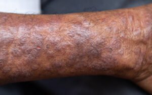
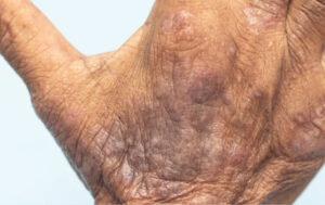
In lighter skin tones, eczema classically presents as erythematous patches or plaques with dryness, excoriation (ie, scratching) and lichenification. However, in people with darker skin tones, visible redness may be far less apparent. Instead, eczema may appear:
- Purplish, dark brown, or ashen grey in appearance
- With prominent hyperpigmentation
- With a tendency towards lichenification (thickened, leathery skin)
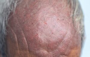
These variations are well described in the literature1,2. It is imperative to recognise that hyperpigmentation is particularly common in skin of colour and that this can be present during active inflammation and persist long after active inflammation has settled1.
Excoriation is universal in eczema and is seen in all skin colours. It is often a very important sign to look for in skin of colour where erythema/redness can be absent.
Hyperpigmentation can cause significant distress, especially when visible areas such as the face or hands are involved. It is important to reassure patients that pigmentation does not represent scarring and that this may fade over time, although it may take months.
Certain clinical patterns may also differ. Papular or follicular eczema is more frequently reported in skin of colour2. Instead of broad erythematous patches, clinicians may observe small, firm papules clustered around hair follicles, particularly on the truncal and extensor surfaces.
Importantly, the absence of obvious redness does not mean the absence of inflammation. Palpation is especially important to note during examination, as features such as warmth, oedema, and thickening may be more reliable indicators of disease activity than colour alone3.
Additionally, clinicians should recognise the psychological impact of dyspigmentation. Hyperpigmentation and hypopigmentation following eczema flares can be particularly distressing in skin of colour and may influence treatment expectations and adherence.
Skin biology
Melanin is the key determinant of skin colour. It is synthesised by melanocytes in the basal layer of the epidermis and is then packaged into melanosomes and transported into surrounding keratinocytes. Skin pigmentation is influenced by the size and distribution of melanosomes. Individuals of African descent have larger melanosomes containing more melanin, which are dispersed throughout the epidermis and take longer to be degraded. Melanocytes produce two types of melanin: eumelanin (the black-brown type) and pheomelanin (the red-yellow type). Individuals with darker skin have a greater proportion of eumelanin, whereas individuals with fairer skin have a greater proportion of pheomelanin4,5.
Diagnostic challenges and severity scoring tools
Many validated eczema severity tools — including EASI (Eczema Area and Severity Index) and SCORAD (Scoring Atopic Dermatitis) — were largely developed and validated in predominantly white populations.
A key limitation is their reliance on visible erythema — red inflamed skin — as a marker of severity. In darker skin tones, erythema may be subtle or masked by background pigmentation3. As a result:
- Severity may be underestimated.
- Disease may be documented as ‘mild’ when symptoms are moderate or severe.
- Patients may not meet thresholds for treatment escalation.
This underestimation can have real-world consequences. If disease severity is under-recognised, patients may be left on insufficient therapy for prolonged periods or may not be referred for systemic treatments6.
For practice and community nurses, this means:
- Place greater emphasis on itch severity, sleep disturbance and quality-of-life impact (eg, NRS Itch score), ask the patient how itchy they have been in last 24 hours on a scale of 1-10
- Assess lichenification, excoriations and extent of body surface area, not just redness.
- Assessing patient-reported outcomes.
- Escalate when disease control is inadequate, even if erythema appears minimal.
Representation in resources: addressing the visual gap
A persistent issue in dermatology education is the underrepresentation of skin of colour in textbooks, image banks and patient leaflets. Many commonly used resources predominantly show eczema on lighter skin tones8.
This visual gap can affect both clinicians and patients:
- Clinicians may feel less confident diagnosing eczema in darker skin.
- Patients may not recognise their condition in educational materials.
- Misdiagnosis (eg, as fungal infection) may occur9.
Improving representation is a shared responsibility. Practitioners can:
- Seek out dermatology image libraries that prioritise diverse skin tones.
- Review patient leaflets before distributing them and consider whether images are representative.
- Signpost patients to reputable online resources that include skin of colour imagery (eg, British Association of Dermatologists, National Eczema Society, and DermNetNZ)10-12
Dr Abi Pararajasingam is a consultant dermatologist and spokesperson for the British Skin Foundation.
Resources
- National Eczema Society. Eczema in skin of colour. https://eczema.org/information-and-advice/types-of-eczema/atopic-eczema/.
- British Association of Dermatologists. Atopic eczema patient information leaflet. https://www.bad.org.uk/pils/atopic-eczema?utm.
- DermNetNZ. Atopic dermatitis. https://dermnetnz.org/topics/atopic-dermatitis.
- Kamp E, et al. Eczema severity scoring in skin of color: a review of current best practice and need for future improvement. J Invest Dermatol 2025; 145(4): 735-748. https://www.jidonline.org/article/S0022-202X(25)00099-5/fulltext#fig1.
References
- Alexis AF, et al. Common dermatologic disorders in skin of color: a comparative practice survey. Cutis. 2007 Nov;80(5):387-94.
- Kaufman BP, et al. Atopic dermatitis in diverse racial and ethnic groups-Variations in epidemiology, genetics, clinical presentation and treatment. Exp Dermatol. 2018;27(4):340-357.
- Zhao CY, et al. The reliability and validity of outcome measures for atopic dermatitis in patients with pigmented skin: A grey area. Int J Womens Dermatol. 2015 6;1(3):150-154.
- Eichenfield LF, et al. Guidelines of care for the management of atopic dermatitis: section 1. Diagnosis and assessment of atopic dermatitis. J Am Acad Dermatol. 2014;70(2):338-51.
- National Institute for Health and Care Excellence (NICE). Atopic eczema in under 12s: diagnosis and management. https://www.nice.org.uk/Guidance/CG57?utm.
- Chiesa Fuxench ZC, et al. Atopic dermatitis in America study: a cross-sectional study examining prevalence and disease burden. J Invest Dermatol. 2019;139(3):583–590.
- Ben-Gashir MA, et al. Quality of life and disease severity are correlated in children with atopic dermatitis. Br J Dermatol. 2004;150(2):284–290.
- Lester JC, et al. Under-representation of skin of colour in dermatology images: not just an educational issue. Br J Dermatol. 2019;180(6):1521–1522.
- Nijhawan, RI, Alexis, AF. Practical approaches to medical and cosmetic dermatology in skin of color patients. 2011. Expert Review of Dermatology, 6(2), 175–187.
- National Eczema Society. Eczema in skin of colour. https://eczema.org/information-and-advice/types-of-eczema/atopic-eczema/.
- British Association of Dermatologists. Atopic eczema patient information leaflet. https://www.bad.org.uk/pils/atopic-eczema?utm.
- DermNetNZ. Atopic dermatitis. https://dermnetnz.org/topics/atopic-dermatitis.
See how our symptom tool can help you make better sense of patient presentations
Click here to search a symptom