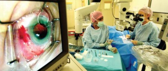
Diabetic retinopathy: an overview

Key learning points:
– Diabetic retinopathy is a chronic and progressive complication of diabetes that threatens sight
– It is classified according to severity, with proliferation of fragile new vessels which are prone to leakage, being a hallmark of poorly controlled disease
– Optimal control of blood sugars and blood pressure has been shown to significantly reduce the risk of progression
Diabetes is a key public health issue. Epidemiological research estimates that in 2010 more than 3.1 million adults in England alone had diabetes and this prevalence is only set to increase. Public Health England estimates that by 2020 8.5% of the population are likely to have diabetes, rising to 9.5% by 2030.1 Visual impairment is the most feared long-term consequence of diabetes2 and the incidence of blindness is 25 times higher in patients with diabetes than in the general population.3 Population screening studies report a baseline incidence of diabetic retinopathy in type 1 diabetes of close to 45%, and in type 2 diabetes estimates range from 25-40%.2,4,5 Indeed, diabetic retinopathy remains the most common cause of blindness in working age people in the UK,3 with profound quality of life and health economic costs.
Having established the importance of understanding diabetic retinopathy, this article aims to provide an accessible overview through considering key ‘what, why, when and how’ questions as a framework for practice as well as understanding the role of primary care nurses in diabetic retinopathy patients.
The ‘what’ and ‘why’
Diabetic retinopathy is a chronic and progressive complication of diabetes that can threaten sight. Chronically elevated blood sugars are known to damage retinal capillaries and alter retinal blood flow. This results in areas of ischaemia and retinal injury. Hypoxic damage ultimately promotes the proliferation of abnormal new vessels that are fragile and prone to leaking and bleeding.
Hypoxia also results in the heightened release of various growth factors that increase vessel permeability and disturb normally tight junctions between cells. As a result, the normal blood/retinal barrier is compromised with subsequent fluid leak/oedema occurring. When this is in the central retina (macula) it is termed macular oedema and can significantly reduce vision.
What does diabetic retinopathy look like?
The changes seen in Table 1, of varying severity, may be seen on fundal examination in diabetic retinopathy.
However, these changes can be difficult to see on fundus examination with a direct ophthalmoscope in community practice, especially if patients are not dilated – this highlights the need for screening photos as a way of community monitoring.
Related Article: Be alert to pancreatitis in patients using GLP-1 weight-loss drugs
How is diabetic retinopathy classified?
Although various slightly different classification systems exist, each system groups patients according
to severity. Broadly there are three main categories to understand:
1. Background or non-proliferative –this group encompasses the majority of patients who have diabetic changes (ie, microaneurysms, haemorrhages, exudates) but who have not yet developed end-stage proliferative changes. As this is such a broad group it is subdivided into mild, moderate or severe changes.2,3
2. Proliferative –this is a late stage of disease characterised by fragile leaky new vessels. The location of the new vessels are specified (ie, new vessels on the disc or elsewhere in the retina) together with the severity (ie, early, high risk, or florid proliferation).
3. Diabetic maculopathy –this specifically refers to retinopathy which affects the macula and therefore threatens central vision, with features such as oedema or ischaemia.
Understanding these broad classifications is important for nurses in practice, both in appreciating the severity of the patients’ eye condition and also in being aware that new visual symptoms such as visual distortion or a drop in visual acuity in diabetic patients could represent changes affecting the macula and warrant further investigation.
It can be useful for nurses to show patients an Amsler chart (see Resources section) as a way of screening for possible macular oedema. This comprises of a grid of straight lines, and patients should be asked whether when they cover either eye, the lines become distorted/are no longer seen as straight.
The complications
Overall, loss of vision from diabetic retinopathy occurs by two mechanisms:
1. Complications of proliferative retinopathy affecting the macula.
2. Loss of peripheral field of vision that results from ischaemia and as a result of laser treatment related damage.2
However, there are many associated complications of diabetic retinopathy that can threaten vision. A permanent gradual reduction in visual acuity can occur due to ischaemia of the central macula region and if leakage occurs (macula oedema) this causes significant distortion and loss of vision. It requires treatment with regular intravitreal injections into the eye in order to limit visual loss and damage to the normal retinal architecture.
Proliferative retinopathy is a hallmark of poor control and has significant consequences. The fine fragile new vessels are prone to acutely bleeding into the eye, known as vitreous haemorrhage, this results in a rapid reduction of vision and can cause a sharp intraocular pressure rise. Dense haemorrhages may necessitate surgical intervention to replace the vitreous gel of the eye. Chronic proliferative disease can also cause tractional pulling on the retina leading to a retinal detachment, which may necessitates difficult vitreoretinal surgery. If patients complain of a sudden drop in vision, practice nurses should seek further help, either consulting a medical practitioner, or if local protocol allows, referring directly to eye casualty to arrange prompt review in the hospital setting.
As well as new vessels proliferating on the retina and optic nerve, prolonged ischaemia can also promote new vessels to proliferate on the iris (surrounding the pupil) and affect the fluid drainage from the front chamber of the eye. This can induce a severe intractable type of glaucoma, giving pain and profound visual loss. We aim to identify and treat all patients before they reach the proliferative stage through the screening programme.

Diagnosing early to prevent complications?
The earlier diabetic retinopathy is diagnosed the sooner measures can be taken to try to limit progression and prevent significant complications. In the UK a highly effective screening service was implemented between 2002 and 2007, offering all diabetic patients access to annual fundal photography. The images obtained are reviewed and graded by specialist teams to detect any retinopathy early and ensure patients are appropriately referred to the hospital eye service in a timely manner.
How is diabetic retinopathy treated?
Reversal of retinopathy is possible in the earlier stages of non-proliferative and prevention is certainly better than cure. The Diabetes Control and Complications Trial (DCCT), the UK Prospective Diabetes Study (UKPDS) and the ACCORD Eye Study have all provided good evidence on the importance of glycaemic and blood pressure control.2,5,6 In the UKPDS, the risk of complications was associated independently and additively with hyperglycaemia and hypertension with significant risk reductions seen with better HbA1c and systolic blood pressure control. A 1% decrement in HbA(1c) equated to a 31% reduction in retinopathy, and a 10mmHg decrement in systolic blood pressure equated to an 11% reduction in photocoagulation or vitreous haemorrhage.5,7
Blood pressure reduction was found to reduce microaneurysms, hard exudates and cotton-wool spots and was associated with less need for photocoagulation. Blood pressure therapy should be initiated aiming for systolic pressures below 130mmHg in those with established retinopathy and/or nephropathy. Blood pressure monitoring should be encouraged in general practice or at home if possible. For diabetics without retinopathy, the normal control target of less than 140mmHg still applies. Lipid-lowering is another approach that may reduce the risk of progression of diabetic retinopathy, particularly macular oedema and exudation. In addition to statins, two large randomised controlled trials of fenofibrate have subsequently confirmed additional benefit in established retinopathy.
Related Article: Low-energy diet improved eating disorder symptoms in patients with type 2 diabetes
Aside from risk modification, no treatment is otherwise indicated for mild background diabetic retinopathy changes. These patients remain on continued monitoring with annual digital photography in the screening programme. However, if there are signs detected in screening of any moderate or severe changes, patients are referred to the hospital eye service for evaluation, with follow up intervals tailored according to the changes seen and likelihood of progression. Patients will be informed that this is an opportunity to tighten their diabetic control and HBA1C monitoring forms an important part of discussions in clinic.
For patients with severe retinopathy and changes approaching the proliferative stage or indeed any signs of new vessels, scatter laser treatment to the retinal periphery (known as PRP) is considered to prevent progression. This treatment aims to reduce the overall retinal oxygen requirements by causing intentional ablation of the outer peripheral retina (not responsible for fine visual acuity). With oxygen demand reduced, there is less ischaemia and consequently less production of growth factors which drive new vessel formation. The principle side effect of PRP laser is peripheral field loss. PRP is a highly effective treatment, shown to reduce the risk of severe visual loss in patients with high-risk characteristics by 50% at two and five years and by up to 70% in moderate risk patients.2
How is diabetic macula oedema treated?
There have been significant recent advances in the treatment of diabetic macular oedema (DMO) with the increasing use of vascular endothelial growth factor (VEGF) inhibitors that are injected into the eye. Anti-VEGF injections stop abnormal blood vessels leaking, growing and bleeding under the retina preventing damage to the retinal light receptors and loss of central vision. Commonly, lucentis (ranibizumab) and eylea (aflibercept) are the drugs used with often very good response and improvement in vision.
Anti-VEGF injections are given after local anaesthetic drops, often by trained nurses, and are generally very safe and cause minimal discomfort. Common side effects patients may experience include a red eye (subconjunctival haemorrhage), floaters or a gritty/sore sensation. Rarely there can be serious complications including a pressure rise, retinal detachment or infection inside the eye. Patients should be informed to contact the eye unit urgently if experiencing reducing vision or significant pain.
The role of the practice nurse
A key requirement for systematic screening is accurate identification in primary care of all those known to have diabetes and the transfer of this information to invite the target population for screening. Practice nurses can certainly help encourage patients to actively engage with screening.
They also have an invaluable role in patient education. We know that progression to more advanced retinopathy is related to the control of diabetes2,8 and its risk can be reduced by intensive blood sugar and blood pressure control. It is therefore important to engage with patients in optimising their diabetic control and approach wider health issues holistically, such as weight, diet and exercise. A personalised HbA1c target should be set in conjunction with the patient and visits should include analysing achievement towards the goal set. Furthermore, practice nurses are in a great position to help ensure co-ordinated care for patients, ensuring they are having regular diabetic reviews and are on appropriate medications. Finally, chronic illness such as diabetes, especially when coupled with sight loss and difficult control, can significantly alter mood and affect quality of life. To be mindful of this is key and practice nurses may also have such a rapport with patients that allows gentle exploration of these issues.
Resources
Amsler chart –macular.org/amsler-chart
References
1. Diabetes Health Intelligence. APHO Public Health Observatories Diabetes Prevalence Model: Key Findings for England, 2010. yhpho.org.uk/resource/view.aspx?RID=81124 (accessed 14 December 2015).
2. The Royal College of Ophthalmologists. Diabetic Retinopathy Guidelines, 2012. www.rcophth.ac.uk/wp-content/uploads/2021/08/2012-SCI-267-Diabetic-Retinopathy-Guidelines-December-2012.pdf
Related Article: Wales diabetes prevention programme cuts risk of developing type 2 diabetes by nearly a quarter
3. Fraser C, D’Amico D. UpToDate. Diabetic retinopathy: Classification and clinical features. uptodate.com/contents/diabetic-retinopathy-classification-and-clinical-features (accessed 14 December 2015).
4. Younis N, Broadbent D, James M, Harding S, Vora J. Current status of screening for diabetic retinopathy in the UK. Diabetic Medicine 2002:19(4):44-49.
5. UK Prospective Diabetes Study Group. Tight blood pressure control and risk of macrovascular and microvascular complications in type 2 diabetes: UKPDS 38. British Medical Journal 1998;317:708-713.
6. The ACCORD Eye Study Group. Effects of Medical Therapies on Retinopathy Progression in Type 2 Diabetes. The New England Journal of Medicine 2010;363:233-244.
7. Kohner EM. Microvascular disease: what does the UKPDS tell us about diabetic retinopathy? Diabetic Medicine 2008;25 Suppl 2:20-4.
8. Amercian Diabetes Association. Diabetic Retinopathy. Diabetes Care 2003, Volume 26, Supplement 1: 100-102.

See how our symptom tool can help you make better sense of patient presentations
Click here to search a symptom


Diabetes is a key public health issue. Epidemiological research estimates that in 2010 more than 3.1 million adults in England alone had diabetes and this prevalence is only set to increase


