Managing leg ulcers in primary care

Key learning points:
- Holistic patient assessment facilitates differential diagnosis in leg ulcer management which enhances patient outcomes
- Compression bandaging remains the ‘gold standard’ for venous leg ulcer management
- The term leg ulcer is not diagnostic, venous, arterial and mixed aetiology remains the three main leg ulcer categories
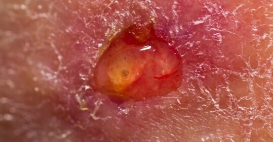
Wounds cost the NHS between £2.3 billion and £3.1 billion annually.1 Among these wounds are leg ulcers. The term leg ulcer is not diagnostic; it describes the situation when the top layer of skin has been lost and the skin beneath is exposed. A holistic assessment is necessary in order to ascertain the cause and underlying pathophysiology and recommend a treatment plan.2,3 Leg ulcers usually fall into one of three categories: venous, arterial and mixed aetiology.4
Venous leg ulcers
A venous leg ulcer is a wound that usually occurs on the medial aspect of the lower leg between the ankle and the knee as a result of venous insufficiency and ambulatory venous hypertension, with little or no progress towards healing within four to six weeks of occurrence.4 The gold standard treatment is compression therapy with the aim of supporting venous return.3,5 It has been noted that approximately 1% of the UK adult population will suffer from a venous leg ulcer at some point in their life, with a prevalence between 0.1% and 0.3%, increasing to 2-3% for patients over the age of 80 and with a higher incidence in women.4,6
Arterial leg ulcers
Arterial leg ulcers are commonly associated with poor blood circulation to the lower leg and foot and are most often due to atherosclerosis.7 With atherosclerosis the arteries become narrowed from deposits of fatty substances, often due to high levels of circulating cholesterol and aggravated by smoking and hypertension. As a result, the arteries fail to deliver oxygen and nutrients to the leg and foot, resulting in tissue breakdown. For those patients, compression therapy is contraindicated and they may require endovascular or surgical revascularisation to promote wound healing.2,3,6
Related Article: ‘Patients not prisoners’: Palliative care nursing behind bars
Mixed aetiology ulcers
Mixed aetiology ulcers are traditionally known as venous leg ulcers with an associated arterial occlusive disease.4,8 However, the term also refers to venous leg ulcers with other contributing factors, such as diabetes, lymphoedema, arthritis, malignancy and many other comorbidities. With changes in societal demographics, mixed aetiology ulcers are likely to be on the increase.9 The current leg ulcer distribution by diagnosis is as shown in table 1, based on a number of studies with different methodology.4 Leg ulcers with a mixed aetiology are usually managed the same way as venous leg ulcer – with compression therapy. However, the level of therapy has to be tailored to each individual.
Diagnostics
To diagnose the category of leg ulcer the clinician must assess a number of factors.
- The patient’s history, which includes: age, occupation or previous employment, personal or family history of skin disorders such as ulceration or dermatitis of the lower limbs. Past medical history, general health, medication, lower leg symptoms, smoking status.
- The appearance of both limbs, observing them for oedema, varicose veins, venous dermatitis, brown staining, atrophie blanche, capillary refill, hair or hairlessness, thickened toenails, dependent rubor.2,3,10
- The nature of the ulcer: site, size, depth, exudate, wound bed composition, clinical signs of infection (heat, inflammation, pain, increased exudate, malodour), signs of dermatitis, wound bed edges (flat, punched out, rolled, circular or jagged edges).2,3,10
- Depending on clinical findings, further investigations may include urinalysis (for sugar, protein, blood); blood tests for albumin, urea and electrolytes, C-reactive protein; swab if there are any clinical signs of infection.2,3,10
It is important that arterial blood flow is measured via ankle brachial pressure index (ABPI).2,3 With advances in technology efficient and cost effective Doppler devices are now available. Regardless of the device used, the formula for calculating ABPI remains ankle systolic pressure divided by brachial systolic pressure.11 This assessment has to be conducted by a trained practitioner. The ABPI assessment will clarify if a patient can be treated with compression bandages or referred to other services. The clinically acceptable Doppler readings are between 0.8-1.3. Other results are summarised in Table 2 below.
However, many clinicians end up fixating on the Doppler number, and ignore the Doppler sounds or wave forms.11 Doppler sounds can be used to determine the level of arterial deterioration. The handheld Doppler machine has three sounds: triphasic, biphasic and monophasic.12 The more advanced ways of calculating ABPIs produce wave forms and notes a recording of whether circulation is normal or abnormal.
Management
Following assessment and differential diagnosis, the clinician has to discuss with the patient the type of therapy that will be effective.13,14
- There are several options and it is essential that clinicians have a clear understanding of:
- The rationale of the therapy, its therapy and characteristics.
- Whether venous return is normal or abnormal.
Properties such as vessel elasticity and stiffness.15
In some clinical settings there are algorithms to support clinicians in decision-making, as demonstrated in figure 1. However, the following recommendations are based on clinical guidance at national level.2,3,16
Management of arterial leg ulcers
The aim of treatment is to enhance arterial blood flow and maintain an effective healing environment. Full assessment including ABPI will determine the arterial element of the leg ulcer. If there is a change in the colour or temperature of the foot, or symptoms suggestive of claudication such as pain in the buttocks when walking short distances, refer for further vascular assessment.3 Patients with an ABPI of less than 0.8 require referral to a vascular team. An ABPI of less than 0.5 requires urgent referral to a vascular team. Management includes:
Related Article: NHSE confirms dates and eligibility for autumn Covid and flu jabs
- Local wound management supported by formulary and clinical expertise.
- Management of surrounding skin to reduce further deterioration.
- Encouraging patients to mobilise to the limits of their capabilities as this will encourage arterial supply.
- Patients should receive healthy lifestyle advice (eg smoking cessation and diet) as appropriate, or referrals to advice services.
- Pain should be assessed using a validated assessment chart and appropriate action taken.
- Compression bandages or compression hosiery are contraindicated.
- Debridement of arterial ulcers is contraindicated in cases of gangrene or stable, dry, ischaemic wounds.
- Dressings should be secured in a non-restricting manner, should not apply compression and should not damage surrounding skin.
Management of mixed aetiology leg ulcer
Treatment aims to increase venous return and maintain an effective healing environment. The degree of arterial insufficiency will dictate whether it is safe to apply compression. There are different types of compression systems which are used in clinical practice, which vary from inelastic to elastic compression bandages, wrap around compression garments, compression stockings, pneumatic pump devices.2,3,4 However, with regards to the management of mixed aetiology leg ulcers the most common way of tailoring compression therapy basing on patient assessment is traditionally done by the use of elastic multilayer bandages.
The vascular team will determine the arterial element and what interventions are required. Reduced compression can be applied for the venous element with the supervision of the vascular team.
Management of venous leg ulcers
The aim of treatment is to improve venous return by increasing velocity flow in the deep veins and to reduce any oedema by decreasing the pressure difference between capillaries and the tissue using compression therapy to promote healing.16 Compression bandaging for venous leg ulcers must aim to deliver 40mmHg by the ankle.2,3,4
Guidelines suggest cleansing of the affected leg should be kept simple, using warm tap water or saline.2
- Irrigate in the shower or bathe in buckets or bowls lined with a clean plastic bag to reduce the risk of cross-infection.
- Gently remove dry skin scales from the legs, particularly around the ulcer edges to allow new growth of epithelium.17
- Soap substitute products should be used to cleanse and moisturise the skin.
In addition to bandaging or compression in general, patients should be encouraged to exercise more than once a day. Exercise should involve the lower leg. Exercise improves the function of the calf pump and improves circulation.
When not exercising, patients should keep their legs elevated as much as is practical. This will help reduce oedema and aids venous drainage from the limb. Patients should be warned that sitting with legs dependent might cause swelling and make the bandages tight and uncomfortable. Sitting in a chair with legs on the foot stool will not help as there will continue to be venous hypertension in the foot, and the position puts additional pressure on the sacrum and buttocks increasing the risk of pressure ulceration. Leg elevation will need to be at the same level as the head in order to facilitate blood circulation.18
Pain
All patients with leg ulceration should be assessed regularly for pain and a management plan formulated to gain acceptable pain control.
- Pain can be a sign of underlying pathology such as arterial disease or infection.
- Exercise and leg elevation may relieve the pain of venous ulcers.
- Compression therapy used for patients with confirmed venous leg ulcers improves pain over time as the ulcer heals. However, pain levels in the first few weeks may rise and patients may require increased analgesia initially.
- Effective analgesia must be tailored to the needs of the patient. Opioid analgesia may be required in some cases.2
Dressing selection
Dressings alone do not heal leg ulcers. Diagnosing and treating the underlying cause is the key to successful treatment.
Allergies may develop at any stage. To reduce the risk of this, avoid where possible: creams, adhesives, topical antibiotics, perfumed lotions and perfumed emollients, paste bandages, lanolin, phenol alcohol and latex gloves.
The type of dressing used on a venous ulcer has little influence on healing when adequate compression is achieved.19 Wound dressings that create and maintain a clean, moist microenvironment are considered optimal for wound healing for basic leg ulcer management.
Related Article: ‘Concerning acceleration’ in drug-resistant gonorrhoea ahead of vaccine programme
Conclusion
Wound care continues to cost the NHS substantial sums of money. Thorough wound assessment will enhance diagnosis, improve patient outcomes and reduce cost. In the management of complex leg ulcers such as arterial ulcer, multidisciplinary working is essential. There are multitudes of dressing products at clinicians’ disposa. However, it is important to have a significant understanding of the rationale of using one product over the other.
References
1. Posnett J, Franks P. The burden of chronic wounds in the UK. Nursing Times 2008;104:44-5.
2. The Royal College of Nursing. The Management of Patients With Venous Leg Ulcers, 2006. rcn.org.uk/__data/assets/pdf_file/0004/107941/001269.pdf (accessed 1 September 2016).
3. Scottish Intercollegiate Guidelines Network. Management of chronic Venous leg ulcers – A National Clinical Guideline, 2010. sign.ac.uk/pdf/sign120.pdf (accessed 1 September 2016).
4. Harding et al. Consensus recommendations: Simplifying venous leg ulcer management, 2015. multimedia.3m.com/mws/media/1082808O/wounds-international-consensus-document.pdf (accessed 1 September 2016).
5. Initiative Chronische Wunden. Recommendations for compression therapy for patients with venous ulcers. Consensus recommendation. EWMA Journal 2013,13:42-7.
6. Gohel MS, Poskitt KR. Chronic ulceration of the leg. Surgery 2010;28:273-6.
7. Anderson I. Aetiology, assessment and management of leg ulcers. Wound Essentials 2006;1:20-37
8. Morison MJ, Ovington LG, Wilkie K (eds). Chronic Wound Care: A Problem Based Learning Approach. Mosby, Edinburgh 2004:164-76.
9. Criqui M, Denenbery J, Bergan J et al. Risk factors for chronic venous disease: the San Diego population study. Journal of Vascular Surgery 2007;46:313-37.
10. Lindsay E, White R (eds). Leg ulcers and problems of the lower limb: A Holistic Approach. Wounds UK, 2008.
11. Vowden P, Vowden K. Doppler assessment and ABPI: Interpretation in the management of leg ulceration. World Wide Wounds, 2001. (accessed 1 September 2016).
12. Donnelly R, Hinwood D, London NJM. Non-invasive methods of arterial and venous assessment. British Medical Journal 2000;320:696-701.
13. Bianchi J, Vowden K, Whittaker J. Chronic oedema made easy. Wounds UK 2012;7(4).
14. Atkin L, Tickle J. A new pathway for lower limb ulceration. Wounds UK 2016;12:32-6.
15. Vowden P. Principles of compression and venous disease: a review. Wounds UK 2014;10:8-10.
16. Clinical Knowledge Summaries. Leg Ulcer – Venous – Management, 2016. cks.nhs.uk/leg_ulcer_venous (accessed 1 September 2016).
17. National Institute for Clinical Excellence. The Debrisoft monofilament debridement pad for use in acute and chronic wounds, Medical Technologies Guidance, 2014. guidance.nice.org.uk/MTG17 (accessed 7 September 2016).
18. Vowden P, Vowden K. How to guide: effective compression therapy. Wound Essentials 2012;7:1-6.
19. Bradely M, Cullum N, Nelson EA, Petticrew M, Sheldon T, Torgerson D. Dressings and topical agents for healing of chronic wounds: a systematic review. Health Technology Assessment 1999;3:1-35.

See how our symptom tool can help you make better sense of patient presentations
Click here to search a symptom


