Supporting vulval health – what a primary care nurse needs to know

In the first of a new mini series, GP and women’s health specialist Dr Louise Clarke provides an overview of vulval health, including how nurses can support patients to understand normal vulval appearance and identify and prevent serious conditions early
Vulval problems are common in primary care. A survey found that over two thirds of GPs were seeing 5 or more women with vulval symptoms a month.1
The most common vulval symptom is itching but women also present with pain, dryness and problems with urinary or sexual function amongst other symptoms.
Women with vulval skin diseases frequently experience misdiagnosis or diagnostic delay. In one study, there was an average 4.6-year diagnostic delay of vulval lichen sclerosus (VLS), the most common vulval skin disease – one patient reported a diagnostic delay of 51 years.2
In addition, vulval complaints can cause wider issues, with increased rates of depression, poor sexual function and decreased work productivity found in women with VLS.3
What is normal?
To identify what is wrong we must be comfortable with what is normal. There is a wide range of normal appearances: all women have outer labia (labia majora), inner labia (labia majora), a clitoris with clitoral hood, a urethral opening and a vaginal opening.
Related Article: Managing weight gain in menopause – what can GPNs do to help?
The appearances often vary from woman to woman; this variation in size, shape and asymmetry is all normal. Many women don’t know their own normal, with 40% of women unable to identify the vagina and 60% unable to identify the vulva on a diagram.4
Pornographic material often features women with hairless, smaller, symmetrical labia and these can be the only other vulvas women have ever seen, if any. As a result, women sometimes present to primary care seeking reassurance that their vulva is normal, or perceiving their normal variation is abnormal. Helpful resources to signpost to include the labia library which has a gallery of diverse normal vulvas and the sexual health charity, Brook, has created a ‘vulva maker’ to emphasise different is normal.
Normal variations in vulval skin
In addition to the diverse appearances of vulval anatomy, there are a few skin changes that could be perceived as abnormal but are benign variants.
- Fordyce spots – these are small (approx. 1-3mm), yellowish smooth bumps or papules on the vulva. They are visible sebaceous glands.
- Vulval papillomatosis – these are tiny (1-2mm across), skin-coloured, shiney finger-like projections. They can occasionally reach up to 6mm in length and are present on the inner labia minora and vestibule of the vagina.
- Angiokeratomas – these are red, purple or blue vascular bumps or papules, 1-5mm in size. They result from a collection of capillaries near the surface of the skin.
- Vulval varicosities – these are varicose veins of the vulva, often appearing in pregnancy. They appear as blue or purple twisted swollen, vessels or lumps. If in pregnancy, they are commonly temporary but can cause pain and itching.
Vulval care
There are many feminine hygiene products available in shops, and none are recommended. Advise patients to avoid all fragranced products such as wipes, perfumes, deodorants, shower gels and fragranced sanitaryware. Emollients can be used to wash with, of which ointments are the most effective, and cotton underwear tends to cause less irritation. A useful leaflet on vulval skincare is available from the British Association of Dermatologists (BAD).
Self-examination
Some vulval skin conditions such as VLS and lichen planus put patients at an increased risk of cancer. Approximately 1400 cases of vulval cancer are diagnosed per year in the UK.5
It is important to recommend that women with these conditions self-examine regularly. I recommend approximately once a month, in line with the University of Nottingham and University of Bristol lichen sclerosis guide. Women should have a system for this so they don’t overlook areas where skin changes could be hidden. For example:
- Get in a comfortable position.
- Use a handheld mirror or mobile phone on a selfie stick to examine – be mindful that recordings may get uploaded to the cloud, there are secure apps available.
- Look around the pubic hair, perineum and anus.
- Spread the outer lips and check all skin folds, particularly around the clitoris.
- Then spread the inner lips, examining these and around the entrance to the vagina.
- It is helpful to look and feel all of the areas.
A comprehensive leaflet and video is available on the British Society for the Study of Vulval Disease website.
Vulval lichen sclerosus
VLS is the most common vulval skin condition, affecting up to 3% of women.6
VLS most frequently affects women of peri or post-menopausal age, but it can be found in all age groups.
Related Article: Picture quiz – genital complaints and knowing when to treat them
Symptoms include itching, burning, pain, dryness and urinary symptoms. If there is diagnostic delay, then VLS can present with irreversible anatomical changes such as fusion of the labia and clitoral hood.
On examination of a patient with VLS, there is typically shiny, white, thin skin found in a figure of 8 distribution around the vulval and peri-anal area (see image). There can be tiny bruises (ecchymoses), thickened areas (hyperkeratosis), fissures and changes to structures.7
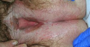
Image: Characteristic appearance of vulval lichen sclerosis, showing thin, shiny white skin around the vulval and perianal area. There are changes to clitoral anatomy and disappearance of the labia minora. The lining of the vagina remains normal.
Women with VLS have an at least 20-fold relative risk of vulval cancer when compared to the general population.8
Early diagnosis and treatment with potent topical corticosteroids reduce the risk of cancer and prevents complications.9 It is crucial that patients understand the increased risk of cancer and the importance of self-examination. The above mentioned lichen sclerosis guide developed by the Universities of Nottingham and Bristol provides a comprehensive resource for patients on all aspects of VLS.
The second article in this series will go into further detail in discussion of common vulval skin conditions.
Related Article: Choosing contraception in women with a family history of thrombosis or cancer
Red flags
Timely referral is important for any worrying lesion that may indicate vulval cancer or the pre-cancerous condition, vulval intraepithelial neoplasia (VIN). Persistent soreness and itching, that doesn’t respond to topical treatments, is a concerning feature. VIN often appears as a raised red, white or pink area.10 An urgent cancer referral is warranted in anyone with an unexplained:
- Lump.
- Ulceration.
- Bleeding.11
Dr Louise Clarke is a GP in Derbyshire an clinical academic at the University of Nottingham. Dr Clarke is the Treasurer of the British Society for the Study of Vulval Disease (BSSVD)
References
- Kandanearachchi P et al. A survey of management of vulvar disorders in the primary health care setting in an urban area of England. Archives of Hellenic Medicine 2018;35(3):405-411
- Cooper S et al. Does treatment of vulvar lichen sclerosus influence its prognosis? Arch Dermatol 2004 Jun;140(6):702-6
- Jabłonowska O et al. Female genital lichen sclerosus is connected with a higher depression rate, decreased sexual quality of life and diminished work productivity. PLoS One 2023 Apr 25;18(4):e0284948
- Eleftheriou-Smith L-M. Half of young women unable to ‘locate vagina’ and 65% find it difficult to say the word. The Independent 2014
- Cancer Research UK. Vulval cancer statistics.
- Leibovitz A et al. Vulvovaginal examinations in elderly nursing home women residents. Arch Gerontol Geriatr 2000 Aug 1;31(1):1-4
- Kirtschig G et al. EuroGuiderm guideline on lichen sclerosus-introduction into lichen sclerosus. J Eur Acad Dermatol Venereol 2024;38(10):1850-73
- Halonen P et al. Lichen sclerosus and risk of cancer. Int J Cancer 2017 May 1;140(9):1998-2002
- Lee A, Fischer G. Diagnosis and Treatment of Vulvar Lichen Sclerosus: An Update for Dermatologists. Am J Clin Dermatol 2018 Oct;19(5):695-706
- British Society for the Study of Vulval Disease. Vulval self-examination for women at increased risk of vulval cancer.
- NICE. CKS. Topics: Gynaecological cancers – recognition and referral. Last revised August 2025
See how our symptom tool can help you make better sense of patient presentations
Click here to search a symptom