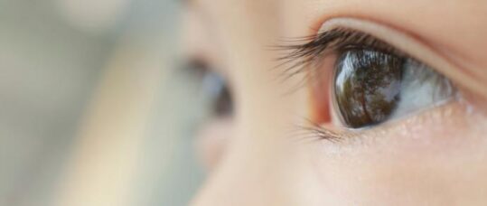
Babies and eyesight problems
Key learning points:
- Babies with low birth weight and come from ethnic minority or deprived socioeconomic groups are most at risk of having eye problems
- It is paramount that the following ocular signs are not missed when assessing a newborn baby: poor fixing and following, abnormal red reflex, photophobia, strabismus and nystagmus
- Certain infections during pregnancy can have an impact on the baby’s eye health
Visual impairment in childhood affects the child’s development, education, and the care given by families and professionals. It also shapes the adult the child becomes, affecting employment and social prospects. The associated life-long burden of disability is substantial and has high economic costs.1
At least four in every 10,000 children born in the UK will be diagnosed as severely visually impaired or blind by their first birthday, increasing to nearly six per 10,000 by the age of 16 years. Children of low birthweight and from ethnic minorities have the highest rates, with those from the most deprived socio-economic groups over-represented. In more than three-quarters of children, visual loss occurs in the context of significant non-ophthalmic impairments or disorders, resulting in death within one year of visual loss in one in 10 most being in the first year of life. The causes of severe visual impairment and blindness are varied and complex, and at least three-quarters of children have disorders that are neither preventable nor treatable, to any degree, with current knowledge.1
Health visitors have a unique opportunity to pick up early signs of poor vision in a newborn baby. Signs that can be manifested by a poorly seeing infant include:
- Poor fixing and following.
- Strabismus (squint).
- Abnormal red reflex.
- Nystagmus / roving eye movements.
- Photophobia.
What is visual acuity? And how is it measured in babies?
Visual acuity is a measure of the central vision. It is the ability to distinguish details ie resolution. It can be measured in different ways. In babies, there are three main methods, namely: visual evoked potentials (which have to be carried out in a hospital setting), preferential looking (acuity cards) and fixation behaviour (can be carried out in the community).2
In fixation behaviour, acuity can be evaluated by a method recognised as CSM:
C stands for ‘central’ – referring to the location of the corneal light reflex as the patient fixates on the examiner’s light monocularly (ie testing one eye at a time). Normally the reflex should be near the centre of the cornea.
S represents ‘steady’ – also referring to the steadiness of fixation on the examiner’s light as it is held motionless and as it is slowly moved about. Ideally this should also be assessed monocularly.2 If a child objects to occlusion of one eye, it is usually a sign that the vision in the uncovered eye is poor.2
M is for ‘maintained’ – relating to the maintenance of fixation when evaluated under binocular conditions (testing both eyes at a time). Inability to maintain fixation, is presumptive evidence of a difference in acuity between the two eyes.2 Therefore, an eye that has eccentric fixation and nystagmoid movements when attempting fixation would have its visual acuity designated uncentral (UC), unsteady (US) and unmaintained (UM).2
Visual development
Normal visual development occurs as a result of both genetic coding as well as experience in a normal visual environment.2
A blink reflex to bright light should be present within several days of birth. The pupillary light reflex is usually present after 31 weeks’ gestation, but it can be difficult to evaluate because of miosis (small pupil) in the newborn. At six weeks of age, the normal baby should be able to make and maintain contact with other human faces and react with facial expressions. At three months, the vision should improve to 6/36; further improving to at least 6/18 by six months.2
Tell tale signs of reduced vision in babies2
Major causes of visual impairment include inherited abnormalities such as cataract, glaucoma and retinal dystrophies; intrauterine insults (eg infection); and acquired problems such as retinopathy of prematurity, and trauma.
Related Article: ‘Concerning acceleration’ in drug-resistant gonorrhoea ahead of vaccine programme
When an infant has not developed good visual attention or ability to fixate and follow objects by the age of three to four months, or objects to occlusion of one eye, further evaluation will need to be undertaken.
Furthermore, disconjugate eye movements may be noted initially (ie the eyes not moving in parallel together), but these should not persist after the age of four months. Wandering/roving eye movements, lack of response to familiar faces and objects and nystagmus all point towards poor visual development. Staring at bright lights and forceful rubbing of the eyes in an otherwise visually disinterested infant are other signs of poor vision that need to be looked out for.
The abnormal red reflex
Reflected light coming from the retina should have an orange-red hue. Leucocoria is an abnormal ‘white’ reflex. There are many different causes for this. The main differential diagnosis that cannot be missed is a Retinoblastoma.
Retinoblastoma is the commonest primary malignant intraocular tumour in childhood. The tumour arises from primitive retinoblasts of the developing retina with loss of function of the Rb tumour suppressor gene (Chromosome 13q14).
The lifetime incidence is one in 15-20,000, and there is no gender or racial predilection. The median age at presentation is less than 12 months in heritable cases, and closer to 24 months in sporadic cases. Presentation after the age of six years is extremely rare.3
Urgent referral (same day, or next day) is vital, and can be life saving
Another cause of leucocoria is cataracts. Examination of the red reflex for the presence of cataract is an essential part of the six week postnatal examination. If abnormality is suspected, prompt referral to an ophthalmologist is needed (within one week).
Eyelid and ‘front of the eye’ disorders2
Anterior segment anomalies such as Ptosis (droopy eyelid especially when covering the pupil), corneal opacities and cataracts result in stimulus deprivation. These can result in amblyopia (lazy eye). In some of these conditions, medical and surgical treatment is available and the amblyopia can be partially or completely reversed.
In these conditions assessment and comparison of the red reflex between the two eyes using a direct ophthalmoscope can give information as to which eye is abnormal.
Back of the eye disorders2
There are many causes of posterior segment anomalies that can lead to reduced visual acuity in infants. The most common are the following:
Leber congenital amaurosis
This is responsible for an estimated 10% of cases of congenital blindness. Infants with this condition may have severe visual impairment at birth, although the visual deficit is noted more typically at two to three months of age with the onset of a coarse nystagmus. The pupillary reactions to light are poor.
Fundus examination is initially normal, although a diffuse pigmentary change, optic nerve pallor, or both may be noted. With time, optic atrophy, narrowing of the retinal vessels and diffuse retinal pigmentary changes occur (similar to retinitis pigmentosa).
Associated neurological abnormalities would include abnormal electrophysiological results, microcephaly, hydrocephaly and seizures. Muscular hypotonia and kidney abnormalities may occur.
Achromatopsia
Is a retinal disorder characterised by total colour blindness. It is inheritied as an autosomal recessive trait. Photophobia, nystagmus and poor visual acuity are common. Fundus examination is usually normal. The electroretinogram (ERG) shows normal scotopic (dark) responses but abnormal photopic (light) responses.
This condition is non-progressive and not associated with other neurologic abnormalities
Infection (particularly TORCH syndrome)
TORCH stands for Toxoplasma, Rubella, Cytomegalovirus (CMV), Herpes Simplex and Syphilis. These congenital infections can lead to marked visual deficit secondary to visual pathway disease and to encephalitis, meningitis, arachnoiditis, optic neuritis and chorioretinitis.
Albinism
Albinism is a group of various conditions that involve the melanin system of the skin, eye or both. The most common form is oculocutaneous albinism.
The major ophthalmic signs are:
- Iris transillumination.
- Foveal hypoplasia or aplasia.
- Photophobia.
- Nystagmus.
- Poor visual acuity.
- High refractive errors.
Albinism patients can have an abnormally large number of crossed fibres at the optic chiasm making their stereopsis (3D vision) poor. Most patients do not have good enough vision to allow them to drive in the future.
Related Article: Action needed to tackle untreated hearing loss in care homes
All forms of this condition are heritable, and genetic counselling is important.
Optic Nerve and Central Nervous System (CNS) anomalies
Anomalies in this category include:
Optic Nerve hypoplasia (ONH)
ONH is characterised by a decreased number of optic nerve axons. The nerve therefore can look small and pale. The condition can be unilateral or bilateral.
The cause of optic nerve hypoplasia is multifactorial. It usually happens as a result of an insult during the first trimester as the optic nerve is developing. Children born to insulin-dependent diabetic mothers are at risk of developing this condition.
Children with ONH often have strabismus (squint) and therefore observation of their visual development is necessary, as amblyopia can develop in these patients; which can be amenable to treatment with patching.
Optic Atrophy
Various inherited and non-inherited conditions can cause optic atrophy. Congenital and acquired forms occur and can be an isolated defect or a part of a systemic disease.
The main primary cause of optic atrophy is:
- Dominant optic atrophy (bilateral visual loss and nystagmus).
Secondary causes of optic atrophy include:
- Hydrocephalus.
- Brain tumours.
- Perinatal hypoxia.
- Central nervous system malformations.
- Trauma (such as Non-accidental injuries).
- Metabolic storage disorders.
Cortical visual impairment (CVI)
This results in varying degree of visual attentiveness. The eye is normal in structure with normal pupillary responses. However, the baby will have ‘searching’ eye movements. CVI may be congenital or acquired. Acquired causes would include pre- or peri-natal intrauterine infection, cerebral dysgenesis, asphyxia, intracranial haemorrhage, hydrocephalus, trauma, meningitis and encephalitis.
Delay in visual maturation
Sometimes, when the eye examination is totally normal, but fixation is poor, the problem is merely delay in visual maturation. Neurological examination can be normal or else the child may have other neurological deficits. Electrophysiological tests are required if no visual improvement happens; otherwise careful monitoring is all that is required.
Coloboma of the optic nerve
Related Article: Government to introduce HPV self-sampling for ‘under-screened’ women
A coloboma is a gap in part of the structure of the eye. It usually affects the inferior aspect of the eye and this corresponds to the lack of closure of the embryonic fissure.
Colobomas of the optic nerve can be isolated or part of a complete chorioretinal coloboma. In this condition, the visual acuity can be mildly or severely affected. It is difficult to predict just on the basis of optic nerve appearance.
Ocular colobomas can be associated with other systemic features which make up the CHARGE syndrome (Coloboma, heart defects, choanal atresia, mental retardation, genitourinary abnormalities and ear abnormalities).
Conclusion
In conclusion, when a baby’s vision is suspected to be sub-normal for age, it is of utmost importance that he/she is referred urgently to the local eye service. Early assessment, investigation and treatment can be key to a more favourable outcome.
References
- Rahi JS, Cable N. Severe visual impairment and blindness in children in the UK. Lancet. 2003 Oct 25;362(9393):1359-65.
- Simon JW, Aaby AA, Drack AV, Hutchinson AK, Olitsky SE, Plager DA, Raab EL, Morse C. Basic and Clinical Science Course Section 6: Pediatric Ophthalmology and Strabismus. San Francisco: American Academy of Ophthalmology; 2007
- Denniston A, Murray P. Oxford Handbook of Ophthalmology. Oxford: Oxford University Press; 2009
See how our symptom tool can help you make better sense of patient presentations
Click here to search a symptom


Visual impairment in childhood affects the child’s development, education, and the care given by families and professionals