A guide to… Doppler ultrasound assessment
Doppler ultrasound assessment is a vital and indispensible element of the process of assessing patients with leg ulceration before planning care.
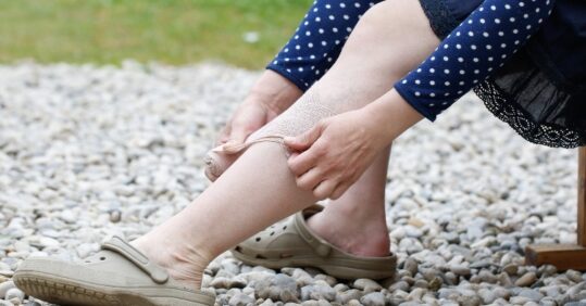
Leg ulceration due to numerous causes (approximately 40) is common, particularly in the over-65 age group (70,000 to 190,000 at any one time in the UK). It is costly to treat (approximately £200m per year) and it often recurs, causing pain and suffering.¹
Most patients are treated in local primary care-based leg ulcer services with specialist services and more complex investigations carried out in secondary care. The Royal College of Nursing (RCN) states: ‘The assessment and clinical investigation of leg ulcer patients should be undertaken by healthcare professionals trained in leg ulcer management’.² Several studies have found a lack of knowledge and wide variation in the nursing management of patients with leg ulceration in terms of assessment.³-⁶
Assessing individuals with leg ulceration
Doppler ultrasound assessment is a vital and indispensible element of the process of assessing patients with leg ulceration prior to planning care; however, evidence shows that many patientassessments do not include Doppler assessment.⁶,⁷ Lack of knowledge, training, equipment, resources and confidence has been cited as reasons for this oversight.⁸ This situation in the past has led to ineffective care and inappropriate treatment.⁴,⁷,⁹
The two potential extremes are that the patient with arterial insufficiency does not receive the correct local management and suffers serious damage and the care of the patient with a chronicvenous leg ulcer is unnecessarily prolonged with the associated risks of extension of the ulcer and infection.
Assessment
The RCN recommends that a thorough clinical history and physical examination should precede patient assessment and investigations to identify any underlying cause and associated disease processes that may be contributing to the development of the ulcer (whether thought to be of venous or non-venous origin) and to aid diagnosis, referrals if necessary, and prognosis.²
Related Article: TikToks promoting ‘dangerous’ sunscreen misinformation attract more engagement
The Scottish Intercollegiate Guideline Network (SIGN) (2010) guidelines stress the importance of calculating the ABPI (ankle brachial pressure index) in patients with chronic venous leg ulceration before treatment begins. The procedure should be carried out by appropriately trained practitioners who should endeavour to maintain their skills.¹⁰
The doppler effect
The term ‘Doppler effect’ dates back to Austrian physicist Christian Doppler (1803-1853) who found that the distance between waves, such as sound or light, changes as an observer of the waves and the source of the waves move relative to each other.
Dopplers detect reflections of small, high frequency sound waves (ultrasound) generated by microscopic vibrations of a ceramic crystal. When ultrasound waves reflect off moving objects such as blood flow, the wave frequency is slightly altered which the Doppler picks up as signals, processes and amplifies so they are audible.
A higher frequency probe (5 or 8MHz) will be suitable for listening to superficial objects such as blood vessels. The transmitted sound wave reflected from moving red blood cells is detected by the receiver and the difference between the frequency of the transmitted sound wave and the frequency of the received signal is known as the Doppler shift. This Doppler shift is typically in the audio range and is converted into the sound heard from the Doppler unit.¹¹
Ankle brachial pressure index (ABPI)
A handheld portable Doppler ultrasound machine is used as a non-invasive method to assess the blood flow in the main arteries of the lower leg carried out using a strict protocol.¹² The procedure of measuring ABPI is described as a ‘simple, practical and objective means of aiding clinical judgement and enables the identification of those patients with a compromised arterial blood supply who would benefit from further vascular assessment’.¹³ It involves calculating the ratio derived from comparing the blood pressure in the upper and lower body; the brachial systolic blood pressure and pedal systolic blood pressure is measured with the patient resting. The ABPI for each leg is calculated by dividing the higher of the ankle pressures by the higher of the twobrachial pressures. The readings should be roughly the same but if the result indicates pressure variation then further investigations may be necessary (see below).
The Doppler assessment should not be viewed as an indicator of, nor does it diagnose, venous disease in the lower leg; however, it will assist and guide management in cases of peripheral arterial disease.¹³ As it is not a diagnostic tool it does help to screen out those patients with any degree of arterial impairment for whom compression therapy would not only be inappropriate but potentially hazardous.¹⁴ It must never be used in isolation but as part of a complete assessment that incorporates the patient’s clinical history and clinical presentation.²,¹⁰ Doppler ultrasound is contraindicated where the patient has had, or is suspected of having, a deep vein thrombosis because of the risk of emboli, cellulitis (because it would be too painful) and severe ischaemia (because of the risk of further tissue damage).
ABPI measurement is an important element in defining a safe level of compression therapy for those patients with venous disease; therefore, it is essential that community nurses perform Doppler ultrasound as an integral part of their assessment.¹²
How to perform doppler assessment/ABPI¹⁵
Equipment
Related Article: Nursing in Practice returns to Scotland with CPD event this September
- Bed or couch.
- Handheld Doppler machine.
- Probe (5 or 8MHz) cleaned with an alcohol-impregnated wipe.
- Transducer gel.
- Sphygmomanometer and cuff.
- Cling film/vapour permeable dressing or equivalent.
- Calculator.
Measuring brachial systolic pressure
- Explain procedure clearly and provide the patient with written information.
- Ensure the environment is warm and comfortable.
- Ask the patient to remove any tight clothing that may interfere with the measurement.
- Ask the patient to lie flat for at least 10 minutes to obtain a resting pressure.¹⁶ If the patient is unable to lie flat, elevate the legs to the level of the heart. By elevating the legs, the hydrostatic pressure to the legs is reduced.
- Measure the brachial systolic blood pressure:
– Select an appropriately sized sphygmomanometer cuff, wrap it around the patient’s upper arm just above the elbow, withthe tubing away from the gel.
– Palpate the brachial pulse and apply ultrasound gel.
– Angle the Doppler probe at 45 degrees to the direction of the
blood flow (towards the heart) and adjust the position to locate
the best signal.
– The Doppler emits an audible signal. Some Doppler ultrasound machines also provide a visual wave form.
– Inflate the sphygmomanometer cuff until the signal disappears, then deflate the cuff slowly and record the pressure at which the signal returns.
- Repeat this procedure using the patient’s other arm.¹⁷
- Use the highest of these two readings to calculate the ABPI.
Measuring ankle systolic pressure
Select an appropriately sized sphygmomanometer cuff, protect fragile skin and the wound. Cover the ulcer with cling film toprevent cross infection and place around the leg just above the ankle and with the tubing away from the gel. Palpate the posterior tibial artery, apply ultrasound gel and locate the best signal.
Inflate the sphygmomanometer cuff until the signal disappears, then deflate the cuff slowly and record the pressure at which the signal returns. Repeat this procedure with either the anterior tibial or peroneal artery.
Repeat for the other leg. Use the highest of these two readings to calculate the ABPI.¹²
The ABPI is calculated using the following equation:
- ABPI = highest ankle systolic pressure/highest brachial systolic pressure; calculate the ABPI for each leg using the formula below or look up the ABPI using a reference chart.
- ABPI = Highest pressure recorded at the ankle for that leg ÷ Highest brachial pressure obtained for both arms.
Vasoconstriction may occur if the patient is uncomfortable, too hot or too cold or anxious, making the sound difficult to hear. In addition, incorrect readings may be a result of inappropriate gel, incorrect size of cuff or probe, incorrect positioning, poor technique and/or inexperience.
Related Article: Lyme disease: what nurses need to know
Interpretation of ABPI
While >1.0 is considered to be normal, it is widely accepted that an ABPI of The holistic assessment should inform the practitioner’s treatment decisions based on other factors; for example, the influence of systemic disease such as diabetes, peripheral vascular disease, rheumatoid arthritis and cardiac failure.¹⁸ In the case of an ABPI between 0.5 and 0.8 and the ulcer diagnosed as being of mixed aetiology, referral to specialist services should be made. An ABPI of
Conclusion
While Doppler ABPI remains an important part of the assessment process it is only a part of the overall holistic assessment of the patient with leg ulceration and should not be used in isolation or as a sole diagnostic tool. Practitioners must be taught how to use the Doppler ultrasound machine competently, remain competent, be able to interpret and document the results correctly, and have the confidence to refer patients to appropriate services in a timely manner.
References
1. Franks P, Moffatt C. Leg ulcers. In: Skin Breakdown: The Silent Epidemic. Hull: The Smith & Nephew Foundation; 2007: 28-32.
2. Royal College of Nursing (RCN). Clinical practice guidelines: The nursing management of patients with venous leg ulcers. London: RCN; 2006.
3. Moffatt C, Franks P. Implementation of a leg ulcer strategy. Br J Dermatol 2004;151:4:857-67.
4. Roe BH, Luker KA, Cullum NA. Assessment, prevention andmonitoring of chronic leg ulcers in the community: report of a survey. J Clin Nurs 1993;2(5):299-306.
5. Bell M. Nurses’ knowledge of the healing process in venous leg ulceration. J Wound Care 1994;3(3):145-50.
6. Elliott E, Russell B, Jaffrey G. Setting a standard for leg ulcer assessment. J Wound Care 1996;5(4):173-5.
7. Stevens J, Franks PJ, Harrington M. A community/hospital leg ulcer service. J Wound Care 1997;6(2):62-8.
8. Schofield J, Flanagan M, Fletcher J, Rotchell L, Thomson S. The provision of leg ulcer services by practice nurses. Nurs Stand 2000;14(26):54-60.
9. Cornwall JV, Dore CJ, Lewis JD. Leg ulcers: epidemiology and aetiology. Br J Surg 1986;73(9):693-6.
10. Scottish Intercollegiate Guideline Network (SIGN). Management of chronic venous leg ulcers. Available from: www.sign.ac.uk/guidelines/fulltext/120/index.html
11. Carefusion. Obstetric and Vascular Dopplers. Product and Accessory Guide. Available from: www.carefusion.com/pdf/Neurology/CF%20SPG%20cat%2011_10_rev14.pdf
12. Vowden KR, Vowden P. Doppler and the ABPI: how good is your understanding? J Wound Care 2001;10(6):197-202.
13. Moffatt C, Martin R, Smithdale R. Leg Ulcer Management. Oxford: Blackwell Publishing; 2007.
14. Jones J. Leg ulceration: correct assessment is vital. Nursing in Practice 2003;9:64-8.
15. Ruff D. Doppler assessment: calculating an ABPI. Nursing Times 2003;99:42:62-5.
16. Yao JST. Pressure measurement in the extremity. In: Bernstein EF (ed). Vascular Diagnosis. St Louis: Mosby; 1993:169-75.
17. Stubbing NJ, Bailey P, Poole M. Protocol for accurate assessment of ABPI in patients with leg ulcers. J Wound Care 1997;6(9):417-8.
18. Marston W, Vowden K. Compression therapy: a guide to safe practice. In: European Wound Management Association (EWMA). Understanding Compression Therapy. London: MEP Ltd; 2003.
19. Stevens J. Diagnosis, assessment and management of mixed aetiology ulcers using reduced compression. J Wound Care 2004;13(8):339-43.
20. Hopkins, A. Leg ulcers: assessment and management plan. Nursing in Practice 2005;25:78-83
See how our symptom tool can help you make better sense of patient presentations
Click here to search a symptom