CPD module: Management of leg ulceration
Julie Hewish, national tissue viability clinical lead, Health Innovation East, offers a guide to the risk factors, presentation, assessment and management of venous and arterial leg ulcers. A longer version of this module is available at Nursing in Practice 365.
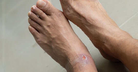
A leg ulcer is defined as an open wound between the knee and just below the ankle joint (malleolus), which has been present for at least two weeks.1 Ulceration is the breakdown of skin, often caused by trauma or surgery. Underlying disease of the vascular system and existing risk factors can mean the healing process for ulcerated skin is challenging.
Approximately 1.5% of the UK adult population are affected by active leg ulceration. This equates to 730,000 people.2 Evidence suggests compression therapy can double the chance of venous ulceration healing within
12 weeks,3 but variation in care practice means only 47% of those affected heal within 12 months.2
Pathophysiology of leg ulceration
Venous leg ulceration
The most common type of ulcer is caused by venous insufficiency and this type accounts for 60-80% of cases.1 It is primarily caused by sustained raised pressure in the veins (venous hypertension), which results in damaged valves and weakens the calf muscle pump action.1
There are multiple risk factors (see Table 1), including deep vein thrombosis (DVT), trauma or lower limb surgery.4
Table 1: Risk factors for venous leg ulceration4
Obesity
Can increase pressure within lower-limb veins
Reduced mobility or walking
Can reduce activation of the calf pump, which helps the blood return to the heart
History of DVT
The obstruction of a blood clot (thrombosis) can damage the valves in the veins
Varicose veins
Vessels walls stretch due to increased pressure in the veins (venous hypertension)
Trauma or surgery to the lower limb
Tensile strength of healed skin is reduced, making the tissue more fragile. Surgery can damage veins and reduce ankle mobility/gait
Increasing age
Reduced mobility, autoimmune diseases such as arthritis, physiological changes in ageing skin
Chronic oedema
Causes inflammatory responses in the skin, can compromise overall skin condition
Family history of leg ulceration
Although leg ulceration is not hereditary, underlying diseases that cause ulceration often are
History of intravenous drug use
Leg ulceration commonly over regular injection sites. Can cause irrevocable damage to vessels
Pregnancy
Can increase pressure on the venous lymphatic system. Lower-leg oedema is common in pregnancy and can cause long-term damage to the vessels and valves
Veins carry deoxygenated blood back to the heart. To prevent backflow (reflux) of blood, most veins have valves that rhythmically close and open to keep the blood moving in one direction and prevent it pooling in the lower limb. When valves are damaged or diseased, they become incompetent, failing to move the blood back to the heart effectively. Increased blood filling in the veins can overstretch the vessel wall so the valves move further apart.4 This gap causes pooling in the blood vessels and leads to symptoms such as lower-limb swelling (oedema) and progressive skin changes such as haemosiderin staining and ankle flare.5
The calf muscle pump is the physical support function for returning venous blood to the heart. Although venous insufficiency is deemed the major cause of lower-limb hypertension, restricted ankle movement can also contribute significantly.6 The pump action relies on patients having a full range of movement in their ankle joint and foot pump. Patients with venous leg ulceration tend to have significantly reduced function in their ankles, which can have a significant impact on quality of life.6
Arterial leg ulceration
Arterial leg ulcers occur due to reduced arterial blood supply to the lower limb. Atherosclerosis, the build-up of fatty deposits (atheroma) in the large vessels, is the most common cause of arterial ulceration.4 As with venous hypertension, damage to and narrowing of the innermost vessel walls (tunica intima) can be caused by sustained raised pressure. This significantly impairs delivery of oxygen and nutrients within skin tissue (tissue hypoxia) leaving the skin vulnerable to ulceration on trauma.7
Related Article: CPD: Gastro-oesophageal reflux disease (GORD) in adults
A number of risk factors, such as smoking, high cholesterol, hypertension and diabetes, can increase
the risk of atheroma. This causes arterial insufficiency and increases the risk of ulceration and/or delayed healing.7
Mixed-aetiology leg ulceration
Mixed-aetiology ulceration applies when there is both venous and arterial disease. Often in these cases, the arterial supply is still enough to perfuse the lower limb but due to the progressive nature and clinical presentation of arterial disease, vascular intervention to resolve arterial insufficiency is often required to prevent ischaemia.7 In practice, this often means treatment decisions for underlying arterial disease will take precedence over those for venous disease. However, in most cases, mild compression can offer relief of venous symptoms in the meantime.2
Lower-limb assessment and ulcer diagnosis
Assessment of the lower limb
All patients with leg ulceration should receive a holistic assessment by a competent health professional within two weeks of presentation to a service.2 The aim of this assessment is to identify underlying risk factors to healing and to diagnose the vascular disease process (aetiology).
A comprehensive lower-limb assessment, along with lifestyle, overall health and relevant medical history, can direct management plans via a collaborative approach with the patient.8 The assessment demands a systematic approach to identify risk factors to healing and avoid longer-term issue. Timely diagnosis of vascular disease is crucial to avoid delayed management.8
The assessment should proceed in the following order:
- The whole person, considering intrinsic and extrinsic risk factors to healing (see Table 1, above).
- The whole limb, looking for skin changes relating to venous or arterial insufficiency5 with an ankle-brachial pressure index (ABPI) reading in order to exclude arterial disease.
- Comprehensive wound assessment to understand wound-bed health and inform a management plan.
During examination of the lower limb:
- Where possible, expose and assess both limbs from foot to upper thigh. Aim to identify any potential issues on the intact limb for preventive measures.
- Identify and document skin changes relating to venous insufficiency.5
- Look for oedema from toes to above the knee joint, which is indicative of venous disease. Note the areas affected and the shape and severity of the swelling, as this will guide compression therapy selection.
- Assess calf muscle strength and tone, and patient mobility and flexibility in the ankle joint.
- Check overall skin condition for the presence of dead skin plaques (hyperkeratosis) across the lower limb, fungal infection and dermatological conditions such as varicose eczema, as these factors will cause further skin deterioration.
- Assess for arterial disease – note and compare changes in colour and temperature of both limbs, working systematically from toes to top of thigh.9
- Observe for inflammation (erythema) and/or pale, cool peripheries (pallor/cyanosis). Be aware of variance in skin presentation in different skin tones.5 Capillary refill is often measured in practice to test arterial perfusion. Arterial disease can affect the results so they should not be judged in isolation.
- Assess for key red flags of peripheral arterial disease (PAD):9
– Does the patient report intermittent pain on exercise or walking, resolving on rest (intermittent claudication)?
– Does the patient report pain at night or at rest, resolving on walking or hanging the limb over the side of the bed (rest pain)?
- If competent to do so, perform a Doppler assessment to establish ABPI.
Doppler assessment
The handheld or automated Doppler assessment is a fundamental element of holistic lower-limb assessment as it helps determine the presence and significance of arterial disease and guides safe compression therapy practice.9 Despite this, it is estimated that in the UK only 15% of patients with leg or foot ulcers have a Doppler ABPI recorded in their notes.10
On completing the Doppler procedure, performing a simple calculation using readings from the ankle and arm provides a ratio figure. Referencing this against a local or national framework of ranges (see Table 3)1 informs the next steps towards compression therapy and/or referrals for specialist vascular assessment.
A normal range for safe, strong compression therapy is often referenced as 0.8-1.3.1 However, obtaining an accurate ABPI can be sometimes be difficult, such as in patients with severe lower-limb oedema or diabetes.11 Results should therefore never be considered in isolation.12 Health professionals should be competent in this procedure and use local protocols and referral pathways.13
Wound assessment
The ulcer should be assessed in a systematic way to identify and address factors within the wound bed environment that could contribute to delayed healing. The assessment should begin with history taking as this will help guide the direction of conversation with the patient.14
Ulcer measurement
The size of the ulcer at four-weekly intervals is seen as a key indicator of progress or deterioration.8,15
A wound surface area reduction of approximately 40% in this time frame is the benchmark for positive progression, with less than 40% being a possible red flag for problematic healing.15 Measurement methods vary in practice but tracing surface area using acetate wound dressings is considered the more accurate.8,15 Wound photography is an important tool as it offers a visual record of wound bed health.
Wound infection
All open wounds will be contaminated with common microorganisms but not all become infected, and most will progress to healing.16 Infection slows healing and increases complications and mortality in patients with leg ulcers.17 Therefore, identifying patients at higher risk of infection, such as the immunosuppressed, is important to reduce further complications.17
The diagnosis of infection in wounds can be challenging. The clinical presentation of local wound infection is categorised into covert (subtle) and overt (classic) symptoms (see Table 2). These may be masked in patients who are immunocompromised and/or have reduced vascular perfusion, as seen in arterial or mixed-aetiology disease.
Table 2: Clinical indicators of local wound bed infection16
Related Article: CPD: Conducting a comprehensive asthma review
Covert (subtle changes at the ulcer bed)
Overt (classic symptoms)
Raised, red granular-looking tissue (hypergranulation)
Redness around the wound (erythema) >2cm
Tissue that looks inflamed (friable) and bleeds easily on cleansing or dressing removal
Local warmth
Increasing exudate in the absence of oedema
Swelling and smelly (purulent), thick exudate
Delayed wound healing beyond expectation
Wound deteriorating despite best practice
Wound swabbing
Wound swabbing determines sensitivities or resistance to medical therapies such as antibiotics when a wound is infected, and cellulitis /systemic infection is present (see Table 2).16 Swabbing is not advocated as routine practice as it can be misleading in a laboratory testing environment. Swabbing the surface of a wound bed may only identify superficial microorganisms without picking up on those in deeper wound tissue (biofilms).16 Clinical judgement based on wound symptoms is a more effective diagnostic tool.12,16
Compression therapy
Venous hypertension is a treatable condition. The National Wound Care Strategy Programme (NWCSP)2 recommends patients be given compression therapy immediately on presentation to prevent longer-term challenges to healing. Strong compression therapy is considered the gold standard for timely healing and improved outcomes.2 Refer to local formularies for guidance on hosiery and bandage systems.
According to the NWCSP, compression therapy is essential as it:
- Controls internal inflammation that causes venous ulceration.
- Reduces and controls the pathophysiology of venous ulceration.
- Helps venous leg ulcers heal more quickly.18
- Can aid pain relief through reducing lower-limb swelling, improving mobility and ulcer bed blood flow.
- Once compression therapy has been deemed safe, the health professional should then assess:
- The level of compression to apply. This depends on the level of compression delivered and product used (see Table 3).12 It is contraindicated in suspected or diagnosed arterial disease. Local protocols should be followed.1
- Choice of compression product should be based on the following considerations:12 the presence of swelling in the lower limb; levels of wound fluid (exudate); lower limb shape and location of ulcer; patient’s height; and psychological and lifestyle considerations.
Table 3: Compression descriptors in line with mmHg delivered12
Mild
Moderate
Strong
Very strong
≤ 20mmHg
20-40mmHg
40-60mmHg
>60mmHg
Compression products
Health professionals should have received appropriate training in therapy selection and be deemed
competent in safe application.2 Manufacturer guidelines must be followed and support sought from industry partners.
- Hosiery compression kits: include two layers, with both delivering up to 40mmHg when applied together. Some kits have a stiffer fabric and are better suited to patients with lower-limb oedema; others have less stiff fabric and are ideal where there is no oedema.3 This system can help promote self-care and reduce ulcer recurrence post healing.
- Bandage compression systems: there is a range of systems, designed to deliver mild to strong compression. They comprise:12,3
– Inelastic/short-stretch bandages
– Elastic/long-stretch bandages
– Multicomponent system (two to four layers)
– Two-layer system.
- Wrap compression garments: these deliver mild-strong compression. Different systems apply depending on lower-limb shape and swelling. They can help promote self-care. There is ongoing research into the effectiveness of these products in venous leg ulcer healing.3
Lower limb skin management
These steps are important for lower limb skin and wound care:2
- Wound bed cleansing and debridement of dead tissue.
- Cleansing of the lower limb to remove wound exudates, dressing residue and dead skin, to encourage emollient uptake.
- Daily emollient therapy routine (where compression regime allows) as per local formulary to maintain the natural skin barrier. Consider barrier products if peri-wound skin is damaged or is vulnerable to damage by exudates.
- Advice on general lifestyle choices, improving nutritional intake, lower-limb exercises and supported self-care.
- Symptom management such as pain relief.
- Providing education and literature to the patient to explain their clinical diagnosis and rationale for compressive therapy.
Author
Julie Hewish is national tissue viability clinical lead, Health Innovation East
Related Article: CPD module: Depression in young men
Find the module
The full CPD module can be found on the Nursing in Practice 365 website.
References
- National Institute for Health and Care Excellence (NICE). Leg ulcer – venous: how should I interpret ankle brachial pressure index (ABPI) results? 2021. Link
- National Wound Care Strategy Programme (NWCSP). Lower limb ulcer recommendations. 2020. Link
- Shi C et al. Compression bandages or stockings versus no compression for treating venous leg ulcers (review). Cochrane Database of Systematic Reviews. 2021; 7: 1-107. Link
- Newton H. Leg Ulcers: Differences between Venous and Arterial. Wound Essentials. 2011; 6; 20-28. Link
- Timmons J and Bianchi J. Disease progression in venous and lymphoevenous disease. The need for early identification and management. Wounds UK. 2011; 4; 3; 59-71. Link
- Chamanga E. Understanding venous Leg ulcers. Community Wound Care. 2018; S6-S15. Link
- Grey J.E., Enoch S., Harding K.G. ABC of Wound Healing. Venous and Arterial Leg Ulcers. BMJ 2006; 332; 347-350. Link
- Mitchell A and Elbourne S. Lower Limb Assessment. British Journal of Nursing. 2020; 29: 1: 18-21. Link
- National Institute for Health and Care Excellence. Peripheral Arterial Disease: diagnosis and management: Clinical Guideline. 2012. Link
- Guest J et al. Cohort study evaluating the burden of wounds to the UKs National Health Service in 2017/2018: Update from 2012/2013. BMJ Open. 2020; 10:e045253. Link
- Elwell R and Sneddon M. Introducing the British Lymphology Society position paper on ankle brachial pressure index. British Journal of Community Nursing. 2019; 24; 5; 207-211. Link
- Wounds UK. Best Practice Statement: Holistic management of venous leg ulceration (second edition). Wounds UK. 2022. Link
- Cain M. et al. Use of ankle-brachial pressure index to assess patient suitability for lower limb compression. Tissue Viability Supplement. British Journal of Nursing. 2022; 31; 20; S6-S14. Link
- Bishop A. Wound Assessment and Dressing Selection: an overview. British Journal of Nursing. 2021; 30; 5. Link
- Nichols E. Wound Assessment Part 1: How to measure a wound. Wound Essentials. 2015; 10; 2; 51-55. Link
- International Wound Infection Institute (IWII) (2022). Wound Infection in Clinical Practice. Wounds International. 2022; 1-57. Link
- Bui U et al. Identifying risk factors associated with infection in patients with chronic leg ulcers. Int. Wound J. 2018; 15; 283-290. Link
- O’Meara S et al. Antibiotics and antiseptics for venous leg ulcers. Cochrane Database Syst.Rev. 2014; 4; 1; Art. No.: CD003557. Link
See how our symptom tool can help you make better sense of patient presentations
Click here to search a symptom