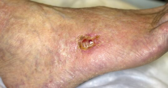
Ten top tips: lower limb wounds

1 Act on red flags
When a patient presents with a wound on the lower limb, assess for ‘red flags’.1 These will require immediate attention from the relevant specialist, in accordance with local pathways. It is important you familiarise yourselves with these.
2 Provide immediate skin and wound care
Cleanse the wound bed, wound edges and the surrounding skin. It is important to encourage the patient to maintain good hygiene of both the leg and foot (especially between the toes) to reduce the risk of infection. Regular application of an emollient will hydrate and protect the skin.
There is no robust evidence to support the use of one type of dressing over another. Therefore, apply a simple, low-adherent dressing with sufficient absorbency, taking into account the latest research evidence as well as patient preferences and cost.2
In the absence of red-flag symptoms (see below), mild graduated compression should be applied for leg ulcers, even where there isn’t any obvious sign of venous disease.1 The benefits of early mild graduated compression are likely to outweigh any risks. Mild graduated compression systems are those that are intended to apply 20mmHg or less at the ankle.1
3 Plan to perform a thorough assessment
A comprehensive assessment should be completed within 14 days for lower-leg wounds.1 For any wound on the foot, a referral should be made to the multidisciplinary foot-care service for assessment and treatment planning within one day,1 in accordance with local pathways.
4 Assessment leads to a diagnosis
A thorough assessment helps to determine the diagnosis and any risk factors for non-healing. It should include consideration of the patient’s clinical and psychological needs, their medication, pain and nutritional intake.1 A comprehensive wound assessment, including photography, will provide a baseline for comparison.
A Doppler ultrasound to calculate the ankle brachial pressure index (ABPI) should be carried out to investigate whether there are problems with arterial blood supply to the lower leg.2 For foot ulcers, toe brachial pressure index (TBPI) measurement can also inform local tissue perfusion. These diagnostic tools should be used as part of
a vascular assessment, which also includes consideration of signs and symptoms of arterial disease. These may include lower-limb hair loss, a change of temperature or colour (pale or blue), or pain experienced in the leg muscles on activity which is relieved by rest.3
A 10g monofilament, or even a simple test such as the ‘touch the toes test’ (otherwise known as the Ipswich Touch Test) can be used to identify any issues the patient might have with neuropathic sensation.1,4
Related Article: Call for regulatory guidelines as NHS adopts AI in dermatology care
5 Ensure the patient understands their condition and things they can do to help
It is very important that the patient understands the underlying cause of their wound so you are able to work together to improve the health of their lower limbs. Information on lifestyle changes to promote healing should include advice on skin care, footwear, exercise and mobility, rest, limb elevation and nutrition.1
Where appropriate, discuss smoking cessation and weight loss. Provide written information, which should explain the treatment plan, including any elements of supported self-care, as agreed with
the patient.
6 Don’t forget analgesia
Lower-limb wounds are often painful so painkillers will be helpful, especially at the start of treatment. When pain is well controlled, patients may be able to tolerate therapy better and participate in supported self-care.
7 Use evidence-based practice for treating venous insufficiency
If diagnosis confirms that a leg ulcer is a result of poor venous return, the patient should be referred to the vascular team for further investigation.5 Modern endovenous interventions are minimally invasive and can speed up ulcer healing and halve the risk of reoccurrence.5,6
If the patient has an adequate blood supply to their lower limb, strong compression therapy should also be applied.1 The recommended therapeutic dose delivers 40mmHg at the ankle.2 The choice
of compression therapy should be made in conjunction with the patient.
8 Refer on for wounds of other or uncertain aetiology
The majority of leg ulcers are caused by venous insufficiency and will be responsive to non-adherent dressings and an adequate dose of compression therapy. But some lower-limb ulcers, including leg ulcers, will be complicated by arterial supply to the limb. A smaller proportion will be due to more unusual conditions, such as skin cancers or other dermatological conditions.
Vascular opinion should be sought wherever there are signs, symptoms and/or confirmation of arterial disease or ischaemia.7 A dermatology referral will be necessary for wounds of other
or uncertain aetiology. In both cases, pending specialist opinion, continue mild graduated compression for leg ulcers if there are no significant symptoms of arterial insufficiency.1
9 Act quickly for foot ulcers
There are many underlying causes of foot ulceration, many of which are associated with an increased risk of amputation.1 This is particularly the case in patients with diabetes, or those with severe peripheral arterial disease, which often manifests as foot ulceration. Despite this, half of all major amputations occur in patients who do not have diabetes.8 Therefore, it is important that there is rapid assessment, diagnosis and treatment planning for all patients with foot ulceration.1 It is also very important to be familiar with your local pathways for foot ulceration and ensure that patients are referred to a multidisciplinary foot service within 24 hours.
10 Review and monitor healing regularly
All lower-limb wounds should be reviewed at each dressing change and at weekly intervals.1 A formal reassessment should take place at four-weekly intervals to monitor the progress towards healing. This should be completed sooner if there is any deterioration, and escalated in accordance with local pathways. If the lower-limb wound remains unhealed at 12 weeks, a comprehensive reassessment should take place and escalated
to the relevant clinical specialist.
Rachael Lee is a clinical lead for the lower limb and surgical wound workstreams of the National Wound Care Strategy Programme. She is predominantly a tissue viability nurse specialist by background, with an interest in wounds research and quality improvement.
Red flags
– Acute infection of leg or foot (eg increasing unilateral redness, swelling, pain, pus, heat).
– Symptoms of sepsis.
Related Article: Abdominal body fat is a higher risk for developing psoriasis
– Acute or chronic limb-threatening ischaemia.
– Suspected deep-vein thrombosis (DVT).
– Suspected skin cancer.
ACTIONS:
– Treat infection.
– Immediately escalate.
– For people in the last few weeks of life, seek input from their other clinicians.
Resources
For further information, see the National Wound Care Strategy Programme’s (NWCSP) Lower Limb Recommendations for Clinical Care. bit.ly/2V6aOmW
The NWCSP’s mission is to implement a consistently high standard of wound care across England by reducing unnecessary variation, improving safety and optimising patient experience and outcomes. Visit the NWCSP website for further information and resources. bit.ly/3DAwMjp
References
1 National Wound Care Strategy Programme, 2020. Recommendations for Lower Limb Ulcers.
2 SIGN. Management of chronic venous leg ulcers: a national clinical guideline. Edinburgh: SIGN, 2010.
Related Article: CPD: Case by case – acute and emergency dermatology presentations
3 NHS, 2019. Overview: peripheral arterial disease.
4 NICE. Diabetic foot problems: prevention and management. London: NICE, 2015.
5 NICE. Varicose veins: diagnosis and management. London: NICE, 2013.
6 Gohel M et al. A randomized trial of early endovenous ablation in venous ulceration. New England Journal of Medicine 2018;378:2105-2114
7 NICE. Peripheral arterial disease. London: NICE, 2018.
8 Ahmad N et al. The prevalence of major lower limb amputation in the diabetic and non-diabetic population of England 2003-2013. Diabetes and Vascular Disease Research 2016;13:348-53

See how our symptom tool can help you make better sense of patient presentations
Click here to search a symptom


